pleural effusion cat ultrasound
Of the 183 cats that received echocardiograms 21 had pericardial effusion with CHF as the primary cause of the pericardial effusion for 86 18 of these cats. Diverse disease processes result in sufficient fluid accumulation within the pleural space to cause respiratory compromise.

Selected Chest Ultrasound Images A Sea Shore Sign Granular Download Scientific Diagram
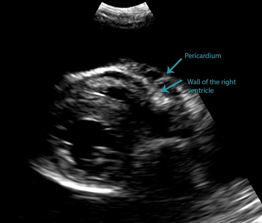
Unlike with a pericardial effusion in the case of accumulation of fluid in the pleural space there is no collapse of the heart walls.

. How the fluid came to be in the pleural space is tied in with this. X-ray and ultrasound imaging of the chest cavity are also very helpful in analyzing the causative factors. Presenting Signs of Pleural Effusion.
Cats presenting with pleural effusion are nearly always in respiratory distress ranging from an increased respiratory rate and effort to open mouth breathing. Pleural effusion in cats with pyothorax in. Conditions which prevent the lungs from fully expanding cause a.
Etiology Prevalence and Epidemiology. Examination of the effusion included determination of specific gravity using a refractometer Atago Company as well as measurement of the total cell count with the Cell-Dyn 3500 System Abott Laboratories. Pleural effusion is the accumulation of fluid in the pleural space resulting from disruption of the homeostatic forces responsible for the movement of pleural fluid.
This non-invasive and quick test can help the veterinarian evaluate the cat quickly. The type of pleural fluid withdrawn will enable your veterinarian to diagnose the cause of the pleural effusion. Approach to pleural effusion in cats.
The most commonly diagnosed cause of pleural effusion in cats is chylothorax. Collection of pleural effusion was performed by blind or ultrasound-guided thoracentesis. Typically this period of remission lasts only 2-9 months and then cats become ill again.
Chest ultrasound has greatly improved the evaluation and interventional management of many pleural diseases. Of the cats that received thoracic ultrasound most exhibited bilateral pleural effusion 93. Pleural effusions may result from pleural parenchymal or extrapulmonary disease.
The therapeutic intervention also provides your first diagnostic test. This review outlines a practical approach to cases of pleural effusion focusing on early recognition and confirmation of pleural space disease stabilisation of the patient and logical diagnostic investigation. 1 Ultrasound has many advantages including the absence of ionizing radiation the sensitivity for detecting small amounts of pleural fluid and the ability to acquire real-time images.
AFAST and TFAST abdominal and thoracic focused assessment with sonography for trauma triage and tracking constitute limited ultrasound examinations that focus on identifying the presence of fluid within the peritoneal pleural and pericardial spaces. Lung ultrasound findings including pleural effusion PLEFF number of Blines and subpleural abnormalities were noted. Reichle J K Wisner E R 2000 Non-cardiac thoracic ultrasound in 75 feline and canine patients.
This can be caused by thoracic lymphangiectasia swollen lymph vessels that leak chyle into the pleural space congestive heart failure obstruction of the cranial vena cava the major vein that returns blood to the heart from the front of the body cancer fungal. Mediastinal lymphoma in cats with feline leukemia carries a poor prognosis with an average survival time of 3 months. Vet Clin North Am Small Anim Pract 30 6 1295-1307 PubMed.
28 Saunders HM V anWinkle TJ Drobatz K. TFAST a standardized and validated thoracic point-of-care ultrasound examination includes 5 acoustic windows. 1999 Peritoneal effusion in cats - 65 cases 1981.
In the below clip from the Sonoscape S2 you can actually see the separation of the right ventricular free wall from the pericardium in a cat. Vet Radiol Ultrasound 41 2 154-162 PubMed. A sample of pleural fluid obtained by piercing the cats chest cavity with a needle will be sent to the laboratory for analysis.
Bilaterally applied chest tube site and pericardial site views plus diaphragmatico-hepatic view also part of AFAST Vet BLUE. Vet Radiol Ultrasound 1998. TFAST is used for rapid detection of pneumothorax and pleural and pericardial effusion.
Padrid P 2000 Canine and feline pleural disease. In cats without feline leukemia mediastinal lymphoma often shows at least a partial response to chemotherapy. There are a lot of causes of pleural effusion in cats transudate or exudate.
Approximately 1 million people develop this abnormality. Blood NTproBNP LUS and FCU evaluating left atrial LA size and presence of pericardial effusion PCEFF were performed in all cats. A restrictive respiratory pattern with increased inspiratory effort is typical.
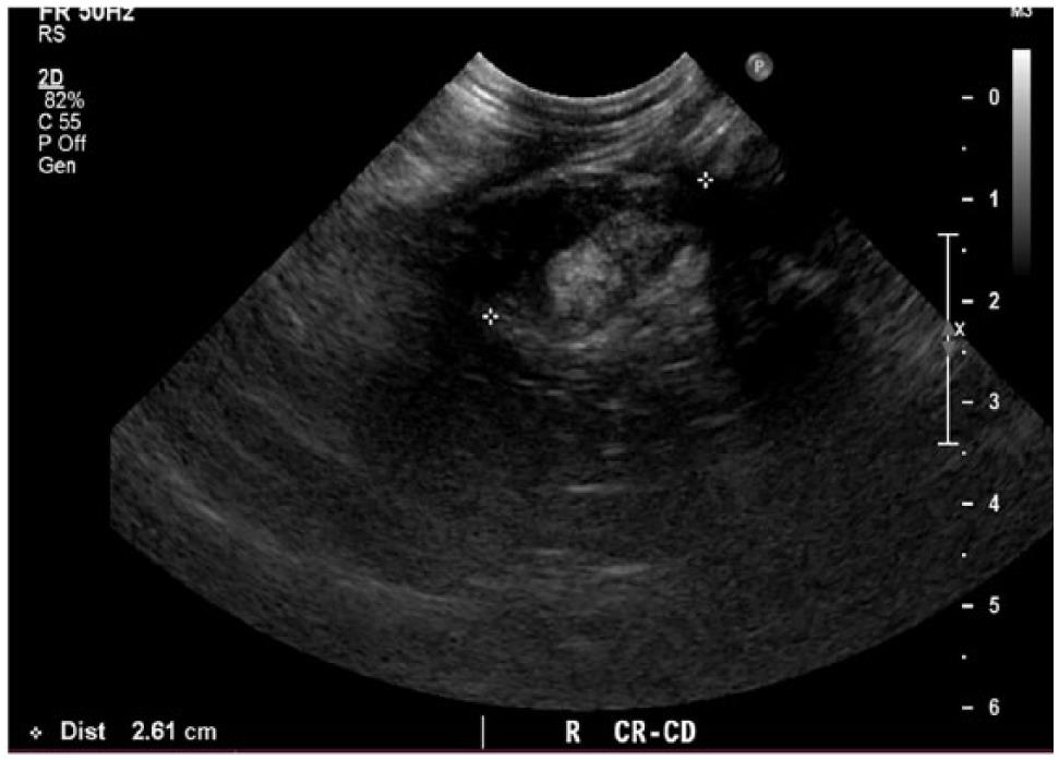
Cats with significant pleural space disease adopt a sternal position with abducted elbows. Thirty-two cats that received thoracic ultrasonography were found to have thoracic masses. A chest ultrasound to look for the presence of fluid within the pleural cavity.
Medical records were evaluated for final diagnosis. Determining the underlying aetiology is key to appropriate management. Vet Radiol Ultrasound 1998.
Rishniw M Weidman J Hornof WJ Hydrothorax secondary to a perinephric pseudocyst in a cat. The rest of the series discusses ultrasound evaluation of specific abdominal organssystems. A restrictive respiratory pattern is rapid and shallow.
To a perinephric pseudocyst in a cat. In the latter situations therapeutic intervention must be initiated quickly to prevent respiratory arrest. This review outlines a practical approach to cases of pleural effusion focusing on early recognition and confirmation of pleural space disease stabilisation of the.
Wright K N Gompf R E DeNovo R C Jr. Pleural effusion is commonly encountered clinically and most patients require.
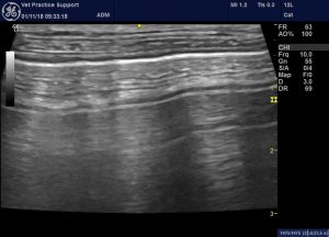
Lung Ultrasound Flooding In Fulminant Pulmonary Oedema In Cats And A Comparison With Pneumonia Vet Practice Support

Pdf Thoracic Ultrasound A Method For The Work Up In Dogs And Cats With Acute Dyspnea Semantic Scholar

How To Ultrasound Detection Of Pleural Fluid Case Study Video Youtube

Top 5 Ultrasound Scenarios In General Practice Clinician S Brief
Spontaneous Cholecystopleural Fistula Leading To Biliothorax And Sepsis In A Cat

Cat Of Figure 1 Thoracic Ultrasound Revealed A Mild Hypoechoic Download Scientific Diagram

Different Types Of Pleural Effusion On Ultrasound Scan A Exudate B Download Scientific Diagram
Veterinary Echocardiography Newsletter 1 Effusions Animal Ultrasound Association

Veterinary Echocardiography Newsletter 1 Effusions Animal Ultrasound Association

Differentiating Pericardial From Pleural Effusion Animal Ultrasound Association
How To Ultrasound Detection Of Pleural Fluid Case Study Video Youtube

Pleural Effusion In A Cat Ultrasound Fip Youtube

Veterinary Echocardiography Newsletter 1 Effusions Animal Ultrasound Association

Large Secundum Asd With Right Sided Enlargement Sonography Student Heart Function Happy People

Pin By Sal Thompson On Radiography Medical Ultrasound Vision Eye Ultrasound

Lung Ultrasound Fundamentals Wet Versus Dry Lung Signs Of Consolidation In Dogs And Cats Veterinary Clinics Small Animal Practice
Animals Free Full Text Lung Ultrasound For Imaging Of B Lines In Dogs And Cats A Prospective Study Investigating Agreement Between Three Types Of Transducers And The Accuracy In Diagnosing Cardiogenic Pulmonary Edema

Use Of Ultrasonography In Veterinary Emergency Rooms Today S Veterinary Nurse

Top 5 Ultrasound Scenarios In General Practice Clinician S Brief